 |
|
||||||||||||
|
|
 |
|
 |
|
 |
 |
|||||||
|
|
 |
|
 |
|
 |
|
|||||||
|
|
|
|
|
|
|
|
|
|
|
|
|
|
|
|
|
|
||||||||||||
|
|
|
|
|
|
|
|
|||||||
|
|
|
|
|
|
|
|
|||||||
|
|
|
|
|
|
|
|
|
|
|
|
|
|
|
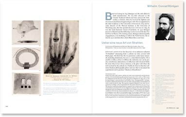
From chapter 67 – Wilhelm Conrad Röntgen
Ueber eine neue Art von Strahlen.
Sitzungb d Würzburger Physik-Med Ges Jahrg 1895. p132.
Würzburg: Verlag der Stahel’schen Buchhandlung; 1896
Röntgen announces his discovery of an unknown radiation “X-Strahlen” emanating from a cathode ray tube covered with black paper. He finds that the ray penetrates easily many materials, including aluminium, but not a 1.5-mm thick lead plate. He is unable to reflect, refract or diffract the radiation, nor can he produce interference phenomena or deflection with electrical fields. He notes that X-rays always emanate at the glass wall where the tube is struck by the cathode ray. Finally, he observes that the radiation blackens photographic plates and uses this to document his experiments and to produce “shadows of handbones”.
CAPTIONS
The famous X-ray picture of the hand of Albert von Köllicker, from the first publication of Röntgen’s discovery.
The equipment used by Wilhelm Röntgen (1845–1923) in his very first experiments with X-rays. Vacuum tubes, an electromagnetic coil employed in his attempts to deflect the X-rays and a plate of lead with apertures for studying the absorption of different materials.
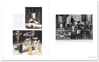
William Coolidge (1873–1975) with his newly developed X-ray tube, which was first used in a clinical setting in 1913. A major advance in X-ray technology, it has a high-vacuum tube with heated tungsten filament as cathode and a tungsten disc as an anode.
Vacuum tubes designed by William Crookes (1832–1919) (at the back of the image) and by Coolidge (in the middle) and by Carl Müller (1845–1912) (front).
Crookes builds vacuum tubes in the last decades of the 19thcentury as part of his investigations of the properties of rarefied gases. Müller starts commercial manufacture of X-ray tubes only months after Röntgen’s discovery.
X-ray treatment in the doctor’s office in 1900.
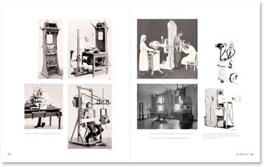
Siemens & Haske X-ray equipment from 1912.
X-ray equipment “Grosscoolinax” from 1933 (at the top).
X-ray room in Caecilien-Krankenhaus Berlin 1935.
X-ray equipment for mass radiography and a mobile X-ray unit (“Pleromobil”) for
operating rooms, both from 1952.